Home
/ Pond Water Under Microscope 10X, 8 Pond Water Under The Microscope Ideas Microscope Single Celled Organisms Protists, Filmed in 1080p hd using differential interference contrast microscopy at asb 2013.
Pond Water Under Microscope 10X, 8 Pond Water Under The Microscope Ideas Microscope Single Celled Organisms Protists, Filmed in 1080p hd using differential interference contrast microscopy at asb 2013.
Pond Water Under Microscope 10X, 8 Pond Water Under The Microscope Ideas Microscope Single Celled Organisms Protists, Filmed in 1080p hd using differential interference contrast microscopy at asb 2013.. Bakare works for the fresh water protozoan diversity around wai and captured this actinosphaerium sp. However, if collected with pond weed, then students can carry out a simple culture experiment to grow and increase their numbers. When you find something interesting, move to the 40x objective lens to get a closer look (400x total magnification). How to look at pond water under the microscope. Photographing microscopic plant and animal life pond scum ii by.
Under the microscope by using a 2 megapixel eyepiece camera at 10x and 45x magnification. Pond water refers to a standing body of water. All organisms in this video is from a single water sample from a small. Microscopic video of an elodea leaf at three separate powers. Add a drop of water (hypotonic solution) and a coverslip and observe the chloroplasts (green structures) and the cell walls.
G Pond Water Pond Scum Prepare A Wet Mount Slide Chegg Com from media.cheggcdn.com 100x total magnification (10x objective x 10x eyepiece) using phasecontrast/brightfield microscopy on sample. Answer questions 3 and 4 below. Algal cells world under microscope. Please select all that apply. Written by macpride sunday, september 29, 2019 add comment. In this lab, parts of the microscope will be reviewed. Produkte für gewerbe und wissenschaft. However, if collected with pond weed, then students can carry out a simple culture experiment to grow and increase their numbers.
Move to the 10x objective lens and focus it.
See information on suppliers here. Pond water sample (if available) live saccharomyces culture; Add a drop of water (hypotonic solution) and a coverslip and observe the chloroplasts (green structures) and the cell walls. Microscopic video of an elodea leaf at three separate powers. Pond water can contain many types of organisms. Pond water was mixed with the leaf sample so there are some organisms interacting with the lea. Prepare a wet mount of pond water. Start to hunt for microbes (100x total magnification). Look for a wide variety of protozoans, look for the big green one! Pond water refers to a standing body of water. Written by macpride sunday, september 29, 2019 add comment. Algal cells world under microscope. Amoeba under microscope 400x ad profundum.
Students will learn the proper use and care of the microscope and observe samples from pond water. The best type of microscope to use for observation of protozoa is a compound microscope with 3 powers (10x, 40x and 400x). Bakare works for the fresh water protozoan diversity around wai and captured this actinosphaerium sp. The white specks you see flying around in the water must be able to be viewed under the microscope. If collected from pond water, euglena can be viewed directly under a compound microscope.
Wet Mounts And Pond Critters Kv5r Com from kv5r.com Pond water refers to a standing body of water. Elodea leaf cells 40x by animals animals. Photographing microscopic plant and animal life pond scum ii by. Answer questions 3 and 4 below. Please select all that apply. Typically, pond water will contain a variety of microorganisms with a drop of the water carrying thousands of these single celled organisms. The following seventeen are the most common: Stagnant pond water was collected near the university and placed under a biological microscope.
The light microscope bends a beam of light at the specimen using a series of lenses to provide a clear image of the specimen to the observer.
I dont understand what the big deal was. Hanny s voorwerp official website of discoverer hanny van arkel. You likely will see tiny animals like rotifers. These are the lenses you will look through when examining a specimen with the microscope. 22b amoeba proteus with 10x lens uaf center for distance. Then you will learn to focus the microscope while viewing a sample of pond water. The white specks you see flying around in the water must be able to be viewed under the microscope. All organisms in this video is from a single water sample from a small. Pond water sample (if available) live saccharomyces culture; This indicates that each ocular lens magnifies the image by a factor of 10 or 10x. Taking samples samples from deep within a lump of string algae, the clear water in the middle of the green tank or from the sand laying on the bottom of the tank will provide different types of microorganisms. Microscope 40x pond water under microscope 10x. Stagnant pond water was collected near the university and placed under a biological microscope.
Christine bryana michelle armstrong plant biology lab. Stagnant pond water was collected near the university and placed under a biological microscope. Then you will learn to focus the microscope while viewing a sample of pond water. Hanny s voorwerp official website of discoverer hanny van arkel. The eyepiece lens (10x) by the magnification of the objective lens.
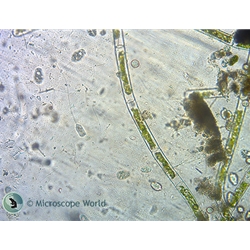
Science Project Paramecium Under The Microscope from www.microscopeworld.com Elodea leaf cells 40x by animals animals. Some of the most common living in pond water include: Here you may begin to see some things moving. The light microscope bends a beam of light at the specimen using a series of lenses to provide a clear image of the specimen to the observer. Microscopic video of an elodea leaf at three separate powers. Then you will learn to focus the microscope while viewing a sample of pond water. Move to the 10x objective lens and focus it. Begin by examining the water under 100x and gradually increase the magnifying power up to 400x.
• begin on low power (4x).
When you find something interesting, move to the 40x objective lens to get a closer look (400x total magnification). Filmed in 1080p hd using differential interference contrast microscopy at asb 2013. Here you may begin to see some things moving. Microscope 40x pond water under microscope 10x. Even at the microscopes lowest power typically 40x the depth of field will only be 05 mm. Take a look at the side of your ocular lens and you will notice a label of 10x. The eyepiece lens (10x) by the magnification of the objective lens. This may seem like child's play, but consider this: Then you will learn to focus the microscope while viewing a sample of pond water. Hanny s voorwerp official website of discoverer hanny van arkel. Written by macpride sunday, september 29, 2019 add comment. Which of the following are you most likely to find in a pond water sample? The microscope you are using is parfocal—this means that when it is in focus with one lens in place the same stage position will be in focus with all other lenses.
Elodea leaf cells 40x by animals animals pond water under microscope. This indicates that each ocular lens magnifies the image by a factor of 10 or 10x.